
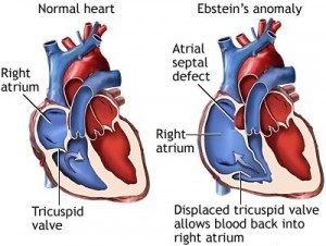
Ebstein’s anomaly presents with a spectrum of congenital abnormalities of the tricuspid valve and the right ventricle.The abnormality of the tricuspid valve leads to tricuspid regurgitation. The degree of tricuspid regurgitation is variable, ranging from mild to severe regurgitation.Presentation is often between the ages of 10 and 30 years but can present at various stages of life:
1. Fetal life: diagnosed incidentally by echocardiography.
2. Neonatal life and infancy: presents with cyanosis and/or severe heart failure. Symptoms presenting in infancy often improve as the pulmonary vascular resistance decreases.
3. Adult life: fatigue, exertional dyspnoea, cyanosis, tricuspid regurgitation and/or right heart failure and palpitations (arrhythmias are common).Additional associated anomalies include bicuspid aortic valves, pulmonary atresia or hypoplastic pulmonary artery, subaortic stenosis, coarctation of the aorta, mitral valve prolapse, accessory mitral valve tissue or muscle bands of the left ventricle, ventricular septal defects and pulmonary stenosis.
Symptoms
1. Cyanosis: common and often due to associated atrial right-to-left shunt and/or severe heart failure. Cyanosis is often transient in neonatal life with recurrence in adult life, but may appear for the first time in adult life. In adult life, cyanosis progressively worsens and may be transiently increased due to paroxysmal arrhythmias.
2. Fatigue and dyspnoea: due to right ventricular failure and decreased left ventricular ejection fraction.
3. Palpitations and sudden cardiac death: due to paroxysmal supraventricular tachycardia or fatal ventricular arrhythmias.
Non-surgical management of Ebsein’s anomaly includes:
1. Antibiotic prophylaxis for infective endocarditis.
2. Management of heart failure.
3. Treatment of arrhythmias.
Surgical
Increasingly the trend is for operative intervention early in the development of heart failure. Ebstein’s anomaly in symptomatic neonates has been shown to be possible, with good survival and excellent functional status.
In patients over 50 years of age with Ebstein’s anomaly, surgery is associated with good long-term survival and improved functional status, although long-term survival might be improved by performing surgery earlier.
1. Tricuspid valve repair is preferred over valve replacement. Bioprosthetic valves are preferred over mechanical prosthetic valves.
2. Both the atrialised portion of the right ventricle and the very dilated, thin-walled right atrium can be resected.
3. Associated heart abnormalities:
– Correction of any associated intra-cardiac defects.
– Associated septal defects may be closed.
– Surgical treatment of associated arrhythmias.
4. Palliative procedures: are usually reserved for severely ill infants with an otherwise poor prognosis. These include:
– Creation of atrial septal defect.
– Closure of tricuspid valve with plication of the right atrium.
– Maintenance of pulmonary blood flow through an aorto-pulmonary shunt.
5. Heart transplantation is appropriate in selected patients.
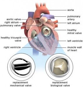
Mitral valve repair is an open heart procedure performed by cardiothoracic surgeons to treat stenosis (narrowing) or regurgitation (leakage) of the mitral valve. The mitral valve lies between the left atrium and left ventricle. The valve has two flaps (cusps). The valve allows blood to flow into the left ventricle when the left atrium contracts. When the left ventricle contracts, the valve closes and the blood flows out through the aortic valve into the aorta. (The aorta is the main artery which takes blood to the body.)
The cusps are stopped from turning inside out by thin strands of tissue called chordae. The chordae (not shown in the diagram) anchor the cusps to the inside wall of the ventricle. The valve or chordae may get damaged or scarred which can prevent the valve from working properly. This can lead to disorders called mitral stenosis, mitral regurgitation, or both.
What is mitral stenosis?
Mitral stenosis means that when the mitral valve opens, it does not open fully. It is narrowed (stenosed) when it is open. So, there is some restriction of blood flow from the left atrium to the left ventricle. This in turn means there is a reduced amount of blood that is pumped out into the body from the left ventricle. In general, the more narrowed the valve, the less blood that can get through, the more severe the problem is likely to be.
What are the causes of mitral stenosis?
– Rheumatic heart disease
This is the cause in most cases. Rheumatic heart disease is a general term which means any heart problem which develops after having an episode of rheumatic fever.
Rheumatic fever is a condition which sometimes follows an infection with a bacterium called the streptococcus. Your body makes antibodies to the bacterium to clear the infection. But, in some people, the antibodies also attack various parts of the body – in particular, the mitral valve. Inflammation of the valve develops which can cause permanent damage and lead to thickening and scarring years late.
– Other causes include:
1. Deposits of calcium (calcification) in parts of the valve. This sometimes occurs in older people.
2. Some congenital (present from birth) heart problems. It is then usually part of a complex heart deformity.
3. Infection of the valve (endocarditis).
4. A complication of various uncommon diseases
Symptoms can include:
1. Shortness of breath. This tends to occur on exercise at first, but occurs at rest if the stenosis becomes worse. This symptom is due to the congestion of blood and fluid in the lungs.
2. Fainting, dizziness or tiredness. If the amount of blood getting through to the ventricle is reduced, the output of blood from the left ventricle to the body is then reduced.
3. Chest pains (angina). This may develop if there is a reduced blood flow to the coronary arteries (the arteries that take blood to the heart muscle).
Chest infections. These are common.
4. Coughing up blood-stained sputum. This may occur due to the congestion of blood and fluid in the lungs.
What are the treatments for mitral stenosis?
Medication
Mild cases may not require any regular medication. Although medicines cannot correct a stenosed mitral valve, some medicines may be prescribed to help ease symptoms, or to help prevent complications.
Cardioversion
Shocking the heart with an electrical current – this is also an option in some people who develop atrial fibrillation as a complication (described earlier).
Surgical treatments
Surgical treatment is needed in more severe cases. There are various options, depending of the exact site and severity of the stenosis.
Stretching the stenosed valve. This is a procedure that does not involve open heart surgery. It is called percutaneous balloon commissurotomy or balloon valvuloplasty. (It is called a commissurotomy, as the area where the valve cusps come into contact with each other are known as the commissures.) This is possible in many cases. It is done by inserting a thin tube called a catheter through the skin (percutaneous) into the main blood vessel in the top of the leg. The catheter is passed up to the heart. The tip of the catheter is placed in the mitral valve opening. A balloon at the tip of the catheter is then inflated to stretch the narrowed valve. This is often successful in widening the narrowed valve.
Valve repair is possible in some cases. This is called mitral commissurotomy or mitral valvotomy. This is usually done by open heart surgery. Basically, the edges (commissures) of valve cusps that have become scarred and fused are shaved back to widen the narrowed valve opening.
Valve replacement is needed in some cases. This may be with a mechanical or a tissue valve. Mechanical valves are made of materials which are not likely to react with your body (for example, those made from titanium), although they can produce a noise which can be heard outside the body. Tissue valves are made from treated animal tissue (for example, valves from a pig).
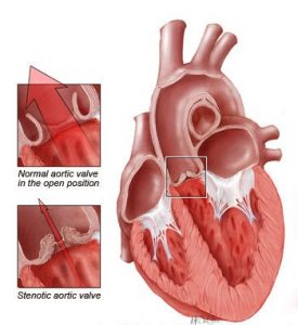
The aortic valve is located between the left ventricle (lower heart pumping chamber) and the aorta, which is the largest artery in the body. Valves maintain one-way blood flow through the heart. Aortic valve disease occurs when the aortic valve does not work correctly. This can be caused by:
Aortic valve stenosis: These stiff, fused, thickened, inflexible valve leaflets lead to the narrowing of the aortic valve, that limits the blood flow. Aortic valve stenosis progresses when calcium is deposited on the valve leaflets, further limiting their mobility. Stenosis can occur in patients with either a tricuspid (3 leaflets) or a bicuspid (2 leaflets) aortic valve.
Aortic valve regurgitation (also called valvular insufficiency, incompetence or “leaky valve”): These valve leaflets do not close completely. Regurgitation causes the blood that is ejected by the heart to immediately flow back into the heart once the heart stops squeezing and relaxes. Regurgitation may occur because of floppy leaflets (prolapse), abnormal congenitally deformed valves (bicuspid or unicuspid), infection of the valve (endocarditis), inability of the leaflets to close tightly due to dilatation of the aorta (aneurysm), holes in the leaflets, or rheumatic valve disease.
Symptoms of aortic valve disease
Many patients with aortic valve disease are asymptomatic (have no symptoms), even when the stenosis (narrowing) or insufficiency (leak) are severe.
Initial symptoms of aortic valve disease usually include:
1. Fatigue
2. Easy tiring
3. Loss of energy
4. Swelling of the ankles
5. Palpitations (extra or skipped heart beats)
More advanced symptoms may include:
1. Shortness of breath
2. Chest pain
3. Dizziness or loss of consciousness
Treatments
Aortic valve repair:While the aortic valve is usually replaced, aortic valve repair may be an option.
Bicuspid aortic valve repair
A bicuspid aortic valve may be repaired by reshaping the aortic valve leaflets allowing the valve to open and close more completely.Bicuspid aortic valve repair may be an option to treat leaking valves, but it can not be used to treat a stenotic or narrowed bicuspid aortic valve.Bicuspid aortic valve repair can be performed using a minimally invasive “J” incision surgical technique. The aortic valve surgery is technically difficult and should be performed by a surgeon with experience repairing aortic valves.
Repair of an enlarged aorta
Aortic valve disease is often associated with enlargement (aneurysm) of the ascending aorta, the initial portion of the aorta (the main blood vessel in the body that originates from the aortic valve).If the enlargement of the aorta is substantial (usually above 4.5 or 5 cm in diameter), this part of the aorta may need to be replaced. The replacement is done at the time of aortic valve repair or replacement. In patients who have leaky aortic valve and an enlarged aorta, a special procedure (David procedure) can be performed
Congenital heart disease (CHD) is the most common congenital disorder in newborns. There are no published data on the occurrence of congenital heart disease in Kerala. However there is general agreement among health workers that it follows a similar pattern all over the world in different geographies. The reported prevalence of CHD at birth ranges from 6 to 13 per 1000 live births. On an average, 8 children are born with congenital heart defects for every 1000 live births in India. Variation is primarily due to the use of different methods to detect CHD (fetal echocardiography versus postnatal referral to a cardiac center). Deriving from the crude birth rate, it…
keep reading

Congenital heart disease (congenital heart defect) is an abnormality in your heart’s structure that you’re born with. Although congenital heart disease is often considered a childhood condition, advances in surgical treatment mean most babies who once died of congenital heart disease survive well into adulthood. While medical advances have improved, many adults with congenital heart disease may not be getting proper follow-up care. If you had a congenital heart defect repaired as an infant, you likely still need care as an adult. Symptoms Symptoms or signs of congenital heart disease may not show up until later in life. They may recur years after you’ve had treatment for a heart defect.…
keep reading
Minimally invasive heart surgery (also called keyhole surgery) is performed through small incisions, sometimes using specialized surgical instruments. The incision used for minimally invasive heart surgery is about 3 to 4 inches instead of the 6- to 8-inch incision required for traditional surgery. The traditional heart surgery procedure Open heart surgery is typically done through a vertical cut placed over the middle of the chest, including full division of the breastbone.While most patients tolerate this well, it can take around 12 weeks or more before the wound is completely healed. This can seriously delay a return to normal activities. These days, it is often possible to avoid such invasive options…
keep readingabout Dr sajan koshy
Pediatric Cardiac Surgeon

Dr Sajan Koshy MS MCh is currently heading the unit of Paediatric & Congenital heart surgery at Aster Medicity. Prior to joining Aster, he was working as senior consultant and head of the department of paediatric cardiac surgery at MIMS Calicut. He started his professional career from alleppey medical college in 1988. He also functioned as a co- convener in first and second symposia on perioperative care of congenital heart disease held in Kochi in the year 2002 and 2004 respectively & as the scientific committee chairman for the national CME of cardio thoracic surgery held at Calicut in September 2007. He has Conducted many lectures in all the districts of North Kerala for the benefit of medical professional bodies to create awareness about congenital heart disease.
read more